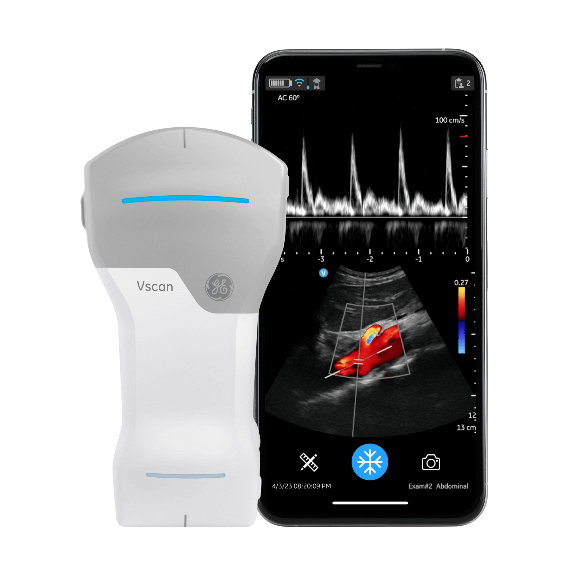
General characteristics
128 physical channel beamforming. Harmonic imaging for increased signal-to-noise ratio and reduced artifacts from side lobes, grating lobes, and reverberations, resulting in superior tissue definition and reduced speckle artifacts. With the greater penetration of lower ultrasound frequencies, high-quality harmonic imaging at greater depth can be performed.
Weight
218 +/- 3 grams, IP67-rated
Drop robustness
MIL-STD-810G, Method 516.7, Table 516.7-7
Scan Time
Start scanning faster with smart connect pairing, which keeps your probe connected to your display device while charging. Total scan time of 50 minutes with fully charged battery (with 80% black and white, 20% color imaging). Recharge battery from 10% to 90% capacity in 75 minutes.
What’s Included in the Standard Vscan offering 123
Vscan Air CL probe, Vscan Air app (Vscan Air for iOS and Vscan Air for Android). Protective carrying case. Wireless charger including USB cable. Country-specific AC adapter. Quick Start Guide. Electronic Instruction Use Guide.
Curved array transducer for deep scanning
Broad-bandwidth curved array: 2 - 5 MHz with center frequency of 3.3 MHz
Number of elements: 128
Footprint: 64 mm x 16 mm (lens)
Viewing angle: Up to 60°
Depth: Up to 24 cm
Specific clinical applications and exam types including: abdominal, fetal/obstetric, gynecological, urological, thoracic/lung, cardiac (adult and pediatric, 40 kg and above), vascular/peripheral vascular, musculoskeletal (conventional), pediatric, interventional guidance (includes free-hand needle/catheter placement, fluid drainage, nerve block, and biopsy)
Linear array transducer for shallow scanning
Broad-bandwidth linear array: 3 - 12 MHz with center frequency of 7.7 MHz
Number of elements: 192
Footprint: 40 mm x 7 mm (lens)
Depth: Up to 8 cm
Specific clinical applications and exam types including: vascular/peripheral vascular, musculoskeletal (conventional and superficial), small organs, thoracic/lung, ophthalmic, pediatric, neonatal cephalic, interventional guidance (includes free-hand needle/catheter placement, fluid drainage, nerve block, vascular access and biopsy)
Color Doppler
Color flow: Color-coded overlay for real-time blood flow imaging
B-mode
Black/white imaging. Black-and-white mode for displaying anatomy in two dimensions in real time
PW
Pulsed wave spectral Doppler displays speed and direction of blood flow allowing velocity measurements
M-mode
Motion mode displays tissue motion over time (along one direction as indicated by m-mode cursor)